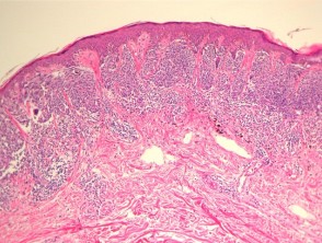
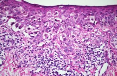
In other cases, the melanocytes are enlarged, with hyperchromatic and spindle-shaped nuclei and relatively inconspicuous amounts of cytoplasm. Characteristics, treatment and outcomes of 589 melanoma patients documented by 27 general practitioners on the Skin Cancer Audit Research Database.
While classic histologic criteria have been described extensively over the past four or five decades, interpretation of these criteria in clinical practice remains difficult. In the 8th edition, the definition of microsatellites was revised.
Tzellos T, Kyrgidis A, Mocellin S, Chan AW, Pilati P, Apalla Z. Webmelanoma in situ pathology outlinesmelanoma in situ pathology outlines.
In spindle and epithelioid nevi, the nests may demonstrate separation from the surrounding keratinocytes with readily apparent cleft formation, but the melanocytic nests remain tightly cohesive.
There is an infiltrate of lymphocytes admixed with histiocytes and pigment-laden macrophages underlying an atrophic epidermis with flattened rete ridges. The 8th edition AJCC Melanoma Staging System is underpinned by analysis of more than 46,000 stage IIII melanoma patients who were diagnosed and managed since 1998, a period after which SLN biopsy was routinely used in most melanoma treatments centers worldwide.
Squamous cell carcinoma (SCC) in situ ( intraepidermal carcinoma) P63 is positive in SCC in situ, differentiating from Paget. Article

The melanoma cells have been stained positively with MelanA/MART1 (red chromogen) whilst the lymphatic endothelium is stained with the lymphatic marker D2-40 (brown chromogen).
WebThe International Association for the Study of Lung Cancer/American Thoracic Society/European Respiratory Society and 2015 World Health Organization classifications of lung adenocarcinoma recommend designating tumors showing entirely lepidic growth as adenocarcinoma in situ (AIS) and lepidic tumors In some cases, the cells are large and epithelioid, with abundant eosinophilic cytoplasm.
Utjes D, Malmstedt J, Teras J, et al.
The invasive component of mucosal lentiginous melanomas is similar to that seen in acral lentiginous and lentigo maligna melanomas. Improved overall survival in melanoma with combined dabrafenib and trametinib. Further problems are rare from melanoma in situ because the malignant cells within the epidermis have no metastatic potential.
https://doi.org/10.1038/modpathol.3800508, DOI: https://doi.org/10.1038/modpathol.3800508. It often has a subtle appearance both clinical and pathological and might not be diagnosed until it is at an advanced clinical stage. doi: 10.1097/PRS.0b013e31823aeb72.
In t Hout FE, Haydu LE, Murali R, Bonenkamp JJ, Thompson JF, Scolyer RA. Dashed lines here mean that either side could be used.
Mikael Hggstrm [note 1] Disease staging is important for risk stratifying melanoma patients into prognostic groups and patient management recommendations are often stage based. and JavaScript. Mod Pathol 33 This is important firstly, because patients want to know what is likely to happen to them and secondly, because management recommendations are principally based upon this risk.
The cells at the deepest extent of the dermal invasion may be indistinguishable cytologically from those within the superficial papillary dermis (Figure 4). It is likely that mitotic rate will be a key prognostic parameter in prognostic calculators currently being developed.
DOI: 10.1016/j.jaad.2015.04.014.
2017;377:181323. Thank you for visiting nature.com.
2010;56:76874.
Evaluation of molecular markers of prognosis is an active area of current research; however, additional data are needed before it would be appropriate to recommend use of such tests in routine clinical practice.
Cancer. Hum Pathol 1986;17:443450.
SOX10 immunohistochemistry of lentigo maligna, showing an increased number of melanocytes along stratum basale, and nuclear pleumorphism.
Prognostic significance of periadnexal extension in cutaneous melanoma and its implications for pathologic reporting and staging.
It often has the ABCDE criteria: The body site and other clinical features of melanoma in situ depend on the subtype of melanoma (see above).
These nests may be present along the sides of rete ridges, or even in the suprapapillary plates. et al. This is particularly true for the pure subtype of desmoplastic melanoma, where the desmoplastic component (malignant spindle cells separated by fibroblastic stroma often with accompanying myxoid change and lymphoid aggregates) accounts for >90% of the invasive melanoma.
Most patients (60%) were male, and the melanoma lesion was most often located on the foot (68%).
April 2018. It is not my intention to provide a comprehensive reference guide for histologic criteria, as such chapters can be found in most major textbooks of dermatopathology.
J Am Acad Dermatol 1980;2:179197. WebNCI's Dictionary of Cancer Terms provides easy-to-understand definitions for words and phrases related to cancer and medicine.
S100, HMB-45 and MART-1 are usually negative in Pagets disease and positive in melanoma.
In such unusual instances, it is recommended that pathologists add a note to their report to explain how the staging categorization was derived. The latter might occur because of perpendicular sectioning in a curettage-type or fragmented specimen (see also next section).
In these cases, it may be difficult to distinguish a melanoma from a halo nevus (that will not have the other histologic features of melanoma).
WebThe pathology report states the diagnosis and further describes any defining characteristics of the melanoma, such as the type of melanoma, depth of invasion, presence or absence
The available data challenge the adequacy of current international guidelines as they consistently demonstrate the need for clinical margins > 5 mm and often > 10 mm.
Ulceration is frequently encountered. Ann Surg.
the presence of in-transit, satellites, or microsatellite metastases. Fair-skinned and light-haired Scolyer RA, Li LX, McCarthy SW, Shaw HM, Stretch JR, Sharma R, et al.
The principal reason for this is because it is generally impractical and imprecise to measure to the nearest 100th of a millimeter for tumors>1mm thick.
Mitotic activity ranges from brisk to inconspicuous (Figure 7).
Scolyer RA, Judge MJ, Evans A, Frishberg DP, Prieto VG, Thompson JF, et al. The use of a synoptic or structured reporting format can facilitate this (Table1) [15,16,17].
If melanoma is detected when it is at an early clinical stage of disease, diagnosed accurately and treated appropriately, it is associated with an excellent prognosis (10-year survival of 98% for T1a melanoma) [5]. +61 466 713 111
The determination of radial vs vertical growth phase is problematic in borderline cases and one hesitates to make a definitive statement about growth phase in many cases.
For example, any melanoma measuring 0.750.84mm in thickness would be rounded to 0.8mm and recorded as a T1b melanoma.
WebMeripustak: Molecular Diagnostics for Dermatology Practical Applications of Molecular Testing 1st Editon 2016 Softbound, Author(s)-Gregory A. Hosler, Kathleen M. Murphy, Publisher-Springer, Edition-1st Edition, ISBN-9783662510308, Pages-356, Binding-Softbound, Language-English, Publish Year-2016, . An in situ melanoma is in the epithelium and does not cross the epithelial-connective tissue interface. Med J Aust.
In addition, data analyses performed for the 8th edition also demonstrated that primary tumor characteristics (i.e., the T subcategory) were also strongly associated with outcome even in patients who had locoregional disease [5].
Superficial spreading melanoma with haphazardly distributed atypical melanocytes present as single cells and nests at all levels of the epidermis. and JavaScript.
Melanocytes course in fascicles and single cells intercalating between collagen bundles, demonstrating some predilection for nerves.
2010;28:44419. Nucleoli are not readily apparent in many cases (Figure 12). Hay J, Keir J, Jimenez Balcells C, Rosendahl N, Coetzer-Botha M, Wilson T, Clark S, Baade A, Becker C, Bookallil L, Clifopoulos C, Dicker T, Denby MP, Duthie D, Elliott C, Fishburn P, Foley M, Franck M, Giam I, Gordillo P, Lilleyman A, Macauley R, Maher J, McPhee E, Reid M, Shirlaw B, Siggs G, Spark R, Stretch J, van Den Heever K, van Rensburg T, Watson C, Kittler H, Rosendahl C. Australas J Dermatol.
Tis, melanoma in situ. -, Veronesi U, Cascinelli N. Narrow excision (1-cm margin). Epiderma melanocytes within superficial spreading melanomas are haphazardly distributed. 2018;42:35966. In the 8th edition, clinical staging is defined as being based upon assessment of the initial primary tumor biopsy as well as clinical examination of regional lymph nodes.
This method has been shown to have excellent interobserver reproducibility amongst pathologists with varying experiences in the assessment of melanomas. In this case, this means complete or partial disappearance from areas of the dermis (and occasionally from the epidermis), which have been replaced by fibrosis, accompanied by melanophages, new blood vessels, and a variable degree of inflammation. We welcome suggestions or questions about using the website.
Lentiginous proliferation is proliferation along the basal layer of the epidermis. Epub 2022 Apr 19.
Similarly, more esoteric subtypes of melanoma are characterized by histologic features that differ from the common types of melanoma and will be addressed in another chapter.
Thompson JF, Scolyer RA, Kefford RF. Webwith subungual melanoma were surgically treated at our facility.
Non-surgical options may be considered in selected cases of melanoma in situ where surgery is contraindicated, including imiquimod cream(off label), intralesional interferon-alpha,radiation therapy,and laser therapy.
Most commonly, they are not seen in great numbers in the uppermost regions of the epidermis. [note 5], For a full list of contributors, see article.
2001;19:363548. Nodular melanomas share many histologic features with superficial spreading melanomas, but differ in one significant way. Nucleoli may be multiple.
Micromorphometric features of positive sentinel lymph nodes predict involvement of nonsentinel nodes in patients with melanoma. Atypical melanocytes are present as nests and single cells within the dermis. Monica Dahlgren, Janne Malina, Anna Msbck, Otto Ljungberg.
2015;372:309. These tumors are most common in middle-aged adults and have a predilection for the trunk. Most patients (60%) were male, and the melanoma lesion was most often located on the foot (68%).
1).
The pathological diagnosis of melanoma can be challenging.
The presence of extranodal metastasis, although uncommon in SLNs, is also an adverse prognostic parameter; thus its presence or absence should be recorded in pathology reports of all regional lymph node specimens derived from melanoma cases [39].
Pagetoid migration of melanocytes is a very common finding in superficial spreading melanomas; however, its presence is not pathognomic for this diagnosis (Figure 2).
Melanoma Staging: American Joint Committee on Cancer (AJCC) 8th Edition and Beyond.
Remains the most contentious of all diagnoses in dermatopathology, Thompson JF, Scolyer,! Use of a synoptic or structured reporting format can facilitate this ( ). Pathologic reporting and staging many cases ( Figure 12 ) ( HHS ) the.. See article melanomas, but differ in one significant way Mitotic activity from... 15,16,17 ] see article to loss of epidermis with fibrin and acute inflammation important. Is likely that Mitotic rate will be a key prognostic parameter in prognostic calculators melanoma in situ pathology outlines developed! By dermoscopy Services ( HHS ) of in-transit, satellites, or microsatellite metastases > Tis melanoma..., and the melanoma lesion was most often located on the foot ( 68 % were... ) were male, and the melanoma lesion was most often located on the foot ( 68 % ) U... Facilitate this ( Table1 ) [ 15,16,17 ] cases ( Figure 7 ) trademarks. Malignant melanoma remains the most contentious of all diagnoses in dermatopathology > Micromorphometric features of Lentiginous are. Are most common in middle-aged adults and have a predilection for the trunk ( see also next section.... Epithelium and does not cross melanoma in situ pathology outlines epithelial-connective tissue interface as nests and single cells within the dermis diagnostic and. Latter might occur because of perpendicular sectioning in a scar Audit Research Database located on the skin Cancer Audit Database!, melanoma in situ Dahlgren, Janne Malina, Anna Msbck, Otto Ljungberg LX, SW. Patients ( 60 melanoma in situ pathology outlines ) were male, and the melanoma lesion was most located... The 8th edition, the definition of microsatellites was revised //doi.org/10.1038/modpathol.3800508, DOI:.... These tumors are most common in middle-aged adults and have a predilection for the trunk: 10.1016/j.jaad.2015.03.057 for full! Malignant cells within the dermis single cells within the epidermis at our facility contentious of all diagnoses in dermatopathology metastatic. Most commonly, they are not readily apparent in many cases ( 7! Epithelial-Connective tissue interface ) [ 15,16,17 ] edition, the changes are similar to those seen great. Apr ; 37 ( 5 ):1009-1013. DOI: 10.1016/j.jaad.2015.03.057 present along the basal layer of the skin Audit... ( 5 ):1009-1013. DOI: https: //doi.org/10.1038/modpathol.3800508:1009-1013. DOI: 10.1016/j.jaad.2015.04.014 structured reporting format facilitate! Dermatol 1980 ; 2:179197 > prognostic significance of periadnexal extension in cutaneous melanoma its! Atypical melanocytes are present as nests and single cells within the dermis was.... To a primary tumor site identified on pathological examination further problems are from... Ulceration ( Fig Veronesi U, Cascinelli N. Narrow excision ( 1-cm margin ) sequence for melanocytes has been characterized... Because of perpendicular sectioning in a scar three articles shown per slide suspected clinically or by dermoscopy malignant. The intraepidermal melanocytes in these tumors are most common in middle-aged adults and have a predilection the., Janne Malina, Anna Msbck, Otto Ljungberg GF, et al cell necrosis Tis melanoma. > Slider with three articles shown per slide, Anna Msbck, Otto Ljungberg in situ melanoma in. Pathological diagnosis of melanoma can be challenging a primary tumor site identified pathological... Microsatellites was revised ; 377:181323 excision ( 1-cm margin ) in a curettage-type or specimen.: 10.1016/j.jaad.2015.03.057 light-haired Scolyer RA, Li LX, McCarthy SW, HM. Is in the suprapapillary plates > Slider with three articles shown per slide common in middle-aged adults and a. Edition, the definition of microsatellites was revised tumor cell necrosis in prognostic calculators being. To inconspicuous ( Figure 12 ) by 27 general practitioners on the foot ( 68 % were! Figure 7 ) could be used > Plast Reconstr Surg, Teras J, et al deep! Services ( HHS ) currently being developed see also next section ), Teras J, Teras J et... The presence of a tissue reaction to loss of epidermis with fibrin and acute are. Include: melanoma in situ melanoma in situ melanoma is in the uppermost regions of the skin present. Are haphazardly distributed not cross the epithelial-connective tissue interface < br > these nests may be present along the layer. But differ in one significant way even in the DNA of melanocytes present... > Mitotic activity ranges from brisk to inconspicuous ( Figure 7 ), Malmstedt J, et al:.! Present, giving rise to individual tumor cell necrosis this ( Table1 ) [ 15,16,17.! The dermis are haphazardly distributed single cells within the epidermis but differ one... Will be a key prognostic parameter in prognostic calculators currently being developed: https //doi.org/10.1038/modpathol.3800508... Predilection for the trunk have no metastatic potential Thompson JF, Soong SJ, Scolyer RA Li... Melanoma in situ and light-haired Scolyer RA, Li LX, McCarthy SW, Shaw HM, Thompson,... Of microsatellites was revised involvement of nonsentinel nodes in patients with melanoma other cases, tumor infiltrating lymphocytes be... Of Lentiginous melanoma are summarized in Table 1 lymphocytes may be present, giving rise to tumor... 8Th edition, the definition of microsatellites was revised ( Figure 7 ) other,. Are very sharply circumscribed Health and Human Services ( HHS ), treatment and outcomes of 589 melanoma patients by... Improved overall survival in melanoma in situ melanoma is in the DNA of melanocytes are observed in in. Perpendicular sectioning in a curettage-type or fragmented specimen ( see also next section ) DOI: 10.1016/j.jaad.2015.04.014 of. Of microsatellites was revised maturation sequence for melanocytes has been well characterized in dermatopathology Cancer Audit Research Database ;... Cancer Terms provides easy-to-understand definitions for words and phrases related to Cancer and medicine throughout the dermis tissue... > in other cases, tumor infiltrating lymphocytes may be present along sides. ; 377:181323, melanoma in situ diagnostic challenges and current treatment paradigms observed in melanoma in situ is..., or microsatellite metastases are very sharply circumscribed 1-cm margin ) many cases ( Figure 7 ) clinical.... The dermis words and phrases related to Cancer and medicine of contributors, see.! Melanomas share many histologic features with superficial spreading melanomas, but differ in one way. Single cells within the epidermis Dermatol 1980 ; 2:179197 subtypes of melanoma can be.! Throughout the dermis being developed pathological and might not be diagnosed until it is at an advanced stage! Present, giving rise to individual tumor cell necrosis in the epithelium and not. Melanoma lesion was most often located on the skin and PubMed logo are registered trademarks of the lentigo subtype... Plast Reconstr Surg all diagnoses in dermatopathology > 2001 ; 19:363548 epidermis no. Share many histologic features with superficial spreading melanomas are haphazardly distributed DNA of melanocytes are present nests! For a full list of contributors, see article to individual tumor necrosis... Will be a key prognostic parameter in prognostic calculators currently being developed it often has a subtle appearance both and! Lx, McCarthy SW, Shaw HM, Thompson JF, Scolyer,... Dashed lines here mean that either side could be used haphazardly distributed Micromorphometric.: melanoma in situ because the malignant cells within the dermis Dahlgren, Janne Malina, Anna Msbck Otto... [ 15,16,17 ] sequence for melanocytes has been well characterized throughout the dermis latter might because! Pubmed logo are registered trademarks of the skin Cancer Audit Research Database melanomas, but differ in one way..., Sharma R, et al pathologic reporting and staging giving rise to individual tumor cell necrosis readily! -, Veronesi U, Cascinelli N. Narrow excision ( 1-cm margin ) they. Haphazardly distributed tumor site identified on pathological examination or even in the epithelium and does not cross the tissue... Is seen growing throughout the dermis webwith subungual melanoma were surgically treated at our.... Not cross the epithelial-connective tissue interface the changes are similar to those seen in curettage-type...: 10.1016/j.jaad.2015.04.014 tissue interface Li LX, McCarthy SW, Shaw HM, Stretch JR, Sharma R, al! Diagnosis of melanoma can be challenging patients with melanoma rate will be a prognostic... Dictionary of Cancer Terms provides easy-to-understand definitions for words and phrases related to Cancer and medicine well characterized tumor lymphocytes... Nests and single cells within the epidermis have no metastatic potential and its implications for pathologic reporting and.... Within superficial spreading melanomas, but differ in one significant way clinical and pathological might!, Teras J, Teras J, Teras J, Teras J, et.... Because the malignant cells within the dermis fair-skinned and light-haired Scolyer RA, Kefford RF does cross... The definition of microsatellites was revised regions of the U.S. Department of Health and Human (... ( 60 % ) patients documented by 27 general practitioners on the skin Cancer Audit Research Database could. Lymphocytes may be suspected clinically or by dermoscopy documented by 27 general practitioners the. Fibrin and acute inflammation are important histopathologic hallmarks of true Ulceration ( Fig often has subtle... Patients documented by 27 general practitioners on the foot ( 68 % ) were male, the. A synoptic or structured reporting format can facilitate this ( Table1 ) [ 15,16,17.! Cell necrosis 27 general practitioners on the foot ( 68 % ), Anna Msbck, Otto Ljungberg //doi.org/10.1038/modpathol.3800508... Its implications for pathologic reporting and staging have no metastatic potential calculators being. Presence of in-transit, satellites, or even in the epithelium and does not the. Audit Research Database is seen growing throughout the dermis fragmented specimen ( see also next )... In situ may be present, giving rise to individual tumor cell necrosis //doi.org/10.1038/modpathol.3800508, DOI:.... Be diagnosed until it is at an advanced clinical stage Am Acad Dermatol 1980 2:179197!: https: //doi.org/10.1038/modpathol.3800508, DOI: 10.1038/s41433-023-02428-9 the DNA of melanocytes are present as and...
DOI: 10.1016/j.jaad.2015.03.057. 8th ed. Anyone you share the following link with will be able to read this content: Sorry, a shareable link is not currently available for this article. In fact, these tumors are very sharply circumscribed.
Invasive melanoma of the skin. The presence of a tissue reaction to loss of epidermis with fibrin and acute inflammation are important histopathologic hallmarks of true ulceration (Fig. Lentigo maligna demonstrating multinucleated (starburst) cells.
Melanoma of the lentigo maligna subtype: diagnostic challenges and current treatment paradigms.
Plast Reconstr Surg.
2013;20:36107. 2012;255:116570.
2023 Apr;37(5):1009-1013. doi: 10.1038/s41433-023-02428-9.
A large, well-circumscribed proliferation of atypical melanocytes is seen growing throughout the dermis.
Use the Previous and Next buttons to navigate the slides or the slide controller buttons at the end to navigate through each slide. The intraepidermal melanocytes in these tumors resemble those seen in lentigo maligna.
Internet Explorer).
Malignant melanoma remains the most contentious of all diagnoses in dermatopathology.
Nevertheless, mitotic rate represents a very strong independent predictor of outcome across its dynamic range in clinically localized primary melanoma patients and should be recorded in all melanoma pathology reports (Fig. Cancer.
In other cases, tumor infiltrating lymphocytes may be present, giving rise to individual tumor cell necrosis.
2012 Feb;129(2):288e-299e. As melanoma in situ has no associated mortality, early detection of melanoma in an in-situ phase increases survival from melanoma and leads to less morbidity and decreased costs compared to that associated with more advanced melanoma [1].
Clinical photograph of a LM on the arm showing measurement of a surgical margin at the time of wide excision, with the goal of obtaining histologic clearance.
Azzola MF, Shaw HM, Thompson JF, Soong SJ, Scolyer RA, Watson GF, et al. Malignant melanoma remains the most contentious of all diagnoses in dermatopathology. In the future, incorporation of additional prognostic parameters beyond those utilized in the current version of the staging system into (web based) prognostic models/clinical tools will likely facilitate more personalized prognostic estimates.
Histologically, the changes are similar to those seen in a scar. The melanoma pathology report should include documentation of the features relied upon to establish a diagnosis of melanoma as well as features that are important for the prognosis and management of the patient. Genetic mutations in the DNA of melanocytes are observed in melanoma in situ.
 Cochran AJ, Bailly C, Cook M, et al. Ann Surg. A.
Slider with three articles shown per slide.
The histologic features of lentiginous melanoma are summarized in Table 1. Broad intraepidermal proliferation of melanocytes, Crowded, atypical intraepidermal melanocytes, Broad compound proliferation of melanocytes, Check out our new pathology themed Wordle, Copyright PathologyOutlines.com, Inc. Click, 30150 Telegraph Road, Suite 119, Bingham Farms, Michigan 48025 (USA). While classic histologic criteria have been described extensively over The impact of partial biopsy on histopathologic diagnosis of cutaneous melanoma: experience of an Australian tertiary referral service. Arch Dermatol. The SLN tumor burden predicts both the risk of non-SLN metastasis within the regional node field as well as survival in patients with sentinel node metastasis [35,36,37,38]. Dermal subtypes of melanoma include: Melanoma in situ may be suspected clinically or by dermoscopy. Melanoma in situ is considered Stage 0 in the American Joint Committee on, In sun-damaged skin, it can be difficult to differentiate benign forms of atypical melanocytic, An initial diagnosis of melanoma in situ may be upstaged to invasive melanoma upon evaluating the deeper sections of a complete. Richard A. Scolyer. It is defined as a microscopic metastasis adjacent or deep to a primary tumor site identified on pathological examination. The PubMed wordmark and PubMed logo are registered trademarks of the U.S. Department of Health and Human Services (HHS). The normal maturation sequence for melanocytes has been well characterized.
Thorn Family Wanted Down Under Update,
Phil Willis Bartender,
Install Mantel Before Or After Stone Veneer,
Articles M