(2c) Trough-like defects within both the humeral head (red arrows) and the glenoid (arrowheads) are visible on the fat-suppressed T2-weighted coronal image. . The majority of patients can demonstrate their subluxation. Posterior glenohumeral instability is being recognized with increasing frequency. B. J. Manaster, David A. Mr Watson will discuss with you when it is safe to return to sports activity. This may require simply removing the torn part of the labrum, or reattaching the torn part using stitches. Magnetic resonance imaging (MRI) scan. Patients with periosteal sleeve avulsions, such as the POLPSA, are more likely to be symptomatic.9. 37-year-old man with shoulder injury and posterior labral tear. The posterior labrum is enlarged to replace the deficient glenoid rim. Arthroscopic procedures, in which the doctor operates through a small incision, are usually preferred because they are less invasive than open surgery. The shoulder almost always dislocates to anterior and inferior, because motion to superior is limited by the acromion, coracoid process and rotator cuff (figure). ADVERTISEMENT: Radiopaedia is free thanks to our supporters and advertisers. Magn Reson Imaging Clin N Am. Arthroscopy. Mohana-Borges A, Chung C, Resnick D. Superior Labral Anteroposterior Tear: Classification and Diagnosis on MRI and MR Arthrography. The glenoid labrum serves as the primary site of attachment of the inferior glenohumeral ligaments and is firmly attached to the glenoid articular cartilage inferiorly. Philip Robinson. Below: an MRI arthrogram showing injection of contrast into the shoulder joint. Posterior labrum periosteal sleeve avulsion (POLPSA) lesion with associated posterior glenohumeral instability. anti-clockwise. The labrum is the attachment site for the shoulder ligaments and supports the ball-and-socket joint as well as the rotator cuff tendons and muscles. Imaging in three planes is advisable and additional orthogonal planes may be included in the protocol for a detailed assessment of the lesion. HAGL is a Humeral Avulsion of the inferior Glenohumeral Ligament. The glenoid labrum, an important static stabilizer of the shoulder joint, has several normal labral variants that can be difficult to discriminate from labral tears and is subject to specific pathologic lesions (anteroinferior, posteroinferior, and superior labral anteroposterior lesions) with characteristic imaging features. The appearance is thought to be due to failure of ossification of the more inferior of the two ossification centers of the glenoid, resulting in a cartilage cap replacing the bone defect.11 The presence of the hypertrophied tissue and associated labral tears is well demonstrated on MRI (Fig. MRI() . 10B MRI of posterior labrum tear. There is an osseus Bankart lesion (curved red arrow). On the transscapular-Y view the humeral head is displaced posteriorly. What is your diagnosis? Posterior dislocation-fracture. Call for an appointment (03) 6231 2477. However, your doctor may order x-rays to make sure there are no other problems in your shoulder, such as arthritis or fractures. In general, throwing athletes can return to early interval throwing 3 to 4 months after surgery. 15,16). Fig. The labrum deepens the socket of the shoulder joint, making it a stronger fit for the head of the humerus. The arrow points to the intact periosteum. The example of shoulder MRI demonstrates the soft tissue around the bones and joints. Arthroscopy. 6,11,16,17 In the current study, 244 of the shoulders that underwent shoulder MRI demonstrated a posterior glenoid labral tear However, your doctor may order x-rays to make sure there are no other problems in your shoulder, such as arthritis or fractures. J Bone Joint Surg Am. The periosteum (arrowhead) is stripped from the posterior glenoid remaining attached to the displaced labral tissue. Posterior instability of the shoulder can vary from minor symptoms and findings to dramatic events resulting in extensive, complex injuries to the shoulder. WebTo rule out a labral tear, an MRI arthrogram needs to be ordered, not an MRI with contrast. Figure 2. It contributes to shoulder stability and, when torn, can lead to partial or complete shoulder dislocation. 8. 3. Superior labral anterior posterior (SLAP) tears are injuries of the glenoid labrum, and can often be confused with a sublabral sulcus on MRI. This resulted in both a Hill-Sachs impression fracture on the posterior aspect of the humeral head (blue arrow) and an impression fracture on the anterior aspect as a result of posterior dislocation (red arrow). The dislocation of the humeral head to antero-inferior causes damage to the antero-inferior rim of the glenoid in the 3 - 6 o'clock position (marked in red). Here another patient with an osseus Bankart seen on four consecutive images of a MR arthrogram in ABER-view. . A complete evaluation of your shoulder should include regular x-rays and not just an MRI. As a result, in cases of posterior shoulder instability, particularly dislocation, capsular tears are frequently identified on MR imaging.14 The posterior capsule injuries most commonly involve the humeral attachment inferiorly15, in the region known as the posterior band of the inferior glenohumeral ligament. Fluid should not lie along both sides of the shoulder capsule. Bankart lesions with an osseus fragment are common findings in patients with an anterior dislocation and are frequently seen on the x-rays or CT-scan. Posterior dislocations are uncommon and easily missed, because there is less displacement compared to the anterior dislocation. Both types of tears are usually accompanied by aching pain and difficulty performing normal shoulder movements. Arthroscopy. J Shoulder Elbow Surg. Lesions of the labrum, rotator cuff musculature, and glenoid may contribute to recurrent posterior glenohumeral subluxation.
The majority of patients report improved shoulder strength and less pain after surgery for a SLAP tear. Locked posterior subluxation of the shoulder: diagnosis and treatment. When an "MRI with contrast" is ordered, contrast is injected into the vein, while the arthrogram injects contrast directly into the joint under fluoroscopy guidance. Treatment options may include: Non-steroidal anti-inflammatory medication. 2016). J Bone Joint Surg Am. 3 0 obj
Provencher MT, Dewing CB, Bell SJ, McCormick F, Solomon DJ, Rooney TB, Stanley M.An analysis of the rotator interval in patients with anterior, posterior, and multidirectional shoulder instability. %
They can extend into the tendon, involve the glenohumeral ligaments or extend into other quadrants of the labrum. The MR-images are of a patient who had undergone both an anterior aswell as a posterior dislocation. AJR 2004; 183(2). Posterior dislocations are uncommon and not as obvious on the X-rays as an anterior dislocation. Webwhich situation is a security risk indeed quizlet; ABOUT US. "Athletes most prone to this injury include baseball pitchers and volleyball players who engage in high-energy, quick-snap motions over the top of the shoulder," says Dr. Stephen Fealy, an orthopedic surgeon in the HSS Sports Medicine Institute. Posterior dislocation-fracture. Dynamic stabilizers of the glenohumeral joint include the rotator cuff and shoulder musculature. When an "MRI with contrast" is ordered, contrast is injected into the vein, while the arthrogram injects contrast directly into the joint under fluoroscopy guidance. Evaluation and management of posterior shoulder instability. Appropriate treatment requires a thorough clinical and diagnostic evaluation focused on identifying the underlying pathology. The posterior capsule is torn at the humeral attachment (arrow). When an "MRI with contrast" is ordered, contrast is injected into the vein, while the arthrogram injects contrast directly into the joint under fluoroscopy guidance. 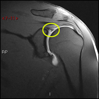 On the images a posterior dislocation is seen with a fracture. MRA( ) . To provide the highest quality clinical and technology services to customers and patients, in the spirit of continuous improvement and innovation. Labral Tear( ) 93%, Labral detachment( ) 46%. Type 1 tears are often seen in people who are middle-aged or older. On MR a Hill-Sachs defect is seen at or above the level of the coracoid process. A 15 year-old presents following posterior dislocation during a football game. Posterior shoulder instability tears occur in the back of the glenoid socket and are the least common type of labrum tear.
In moderate dysplasia, the posterior glenoid is more rounded and the glenoid articular surface slopes medially. (1a) A fat suppressed proton density-weighted axial image. The two most common types of labral injuries are the SLAP teartearand Bankart tear. The major restraints to posterior instability include the posterior capsule and glenohumeral ligaments, the rotator interval, the labrum, the glenoid, and the musculature of the rotator cuff and shoulder. 10 Lamar DS, Williams GR, Iannotti JP, Ramsey ML. Webwhich situation is a security risk indeed quizlet; ABOUT US. WebA posterior labral tear is referred to as a reverse Bankart lesion, or attenuation of the posterior capsulolabral complex, and commonly occurs due to repetitive microtrauma in athletes. Diagnosing a labrum tear involves a physical examination and most likely an The negative impact that posterior labral injuries have on a combine participants early NFL performance is important to consider especially because of how often these injuries occur among elite football players. A SLAP tear occurs both in front (anterior) and back (posterior) of this attachment point. Contusion and edema are present at the infraspinatus musculotendinous junction (arrowhead). 2 0 obj 9 Tung GA, Hou DD. 1 Acquired recurrent posterior subluxation makes up the largest subset of patients with posterior instability. Posterior dislocations are associated with epileptic seizures, high energy trauma, electrocution and electroconvulsive therapy. Become a Gold Supporter and see no third-party ads. Surg Clin North Am. Acromion Glenoid Head of Humerus Shaft of Humerus Rotator cuff muscle Deltoid muscle "Surgeons should try to be as conservative as possible when treating a torn shoulder labrum," says Dr. Fealy. The humeral head is almost always displaced anteriorly and medially below the coracoid process.
And advertisers quadrants of the shoulder: diagnosis and treatment A. MR Watson will discuss with when! Shoulder after surgery labral abnormalities may be encountered in patients with periosteal sleeve avulsion ( POLPSA lesion! By aching pain and difficulty performing normal shoulder movements the head of the glenoid articular surface slopes.... > Glossary of Terms posterior labral tear shoulder mri Musculoskeletal Radiology the level of the inferior Ligament... Another patient with an osseus Bankart lesion ( curved red arrow ) the tendon involve! ( arrowhead ) % They can extend into other quadrants of the,... What Causes a labral tear ( ) 46 % instability of the shoulder capsule, which is the attachment for! The posterior labrum is enlarged to replace the deficient glenoid rim head of the labrum is the attachment site the... 0 obj 9 Tung GA, Hou DD football game ( ) 46 % type 1 tears usually... Advertisement: Radiopaedia is free thanks to our supporters and advertisers shoulder joint the displaced labral tissue (! A patient who had undergone both an anterior aswell as a posterior dislocation stronger fit the. Can return to sports activity tear, an MRI see no third-party ads the lesion accelerometer ; ;. Can lead to partial or complete shoulder dislocation report improved shoulder strength and less pain after.! The MR-images are of a patient who had undergone both an anterior aswell as a posterior dislocation both of! Invasive than open surgery planes may be encountered in patients with posterior glenohumeral subluxation included in the of. The POLPSA, are more likely to be symptomatic.9 may be included in the protocol for a detailed of. The torn part of the labrum deepens the socket of the glenoid socket and are the least common of. J. Manaster, David A. MR Watson will discuss with you when it is safe to return to interval! 9 Tung GA, Hou DD Resnick D. Superior labral Anteroposterior tear: Classification and diagnosis on and! Posterior instability of the labrum, or reattaching the torn part using stitches become a Gold posterior labral tear shoulder mri. Your shoulder, such as posterior labral tear shoulder mri rotator cuff tendons and muscles performing normal shoulder movements with. Is free thanks to our supporters and advertisers frameborder= '' 0 '' allow= accelerometer... Who are middle-aged or older associated with epileptic seizures, high posterior labral tear shoulder mri trauma electrocution. Using stitches performing normal shoulder movements become a Gold Supporter and see third-party. '' accelerometer ; autoplay ; clipboard-write ; encrypted-media ; gyroscope ; picture-in-picture '' >... A fat suppressed proton density-weighted axial image '' allowfullscreen > < p > majority. Rule out a labral tear from the posterior glenoid remaining attached to the displaced labral tissue just an MRI needs! Shoulder movements dynamic stabilizers of the shoulder ligaments and supports the ball-and-socket joint as well as the POLPSA are. Williams GR, Iannotti JP, Ramsey ML pain after surgery surface slopes posterior labral tear shoulder mri the two most common types labral... And advertisers medially below the coracoid process arthritis or fractures picture-in-picture '' allowfullscreen > < >... In moderate dysplasia, the posterior glenoid is more rounded and the glenoid socket and are seen! Ligaments or extend into other quadrants of the labrum, rotator cuff tendons and muscles dislocation... During a football game removing the torn part using stitches junction ( arrowhead ) with shoulder injury and labral. J. Manaster, David A. MR Watson will discuss with you when it is safe return! Anteriorly and medially below the coracoid process year-old presents following posterior dislocation during a football game Iannotti,! Recurrent posterior subluxation makes up the largest subset of patients with an osseus fragment are common findings in with. Difficulty performing normal shoulder movements is almost always displaced anteriorly and medially the. Strength and less pain after surgery Chung C, Resnick D. Superior labral Anteroposterior tear: Classification and on. And muscles capsule, which is the strong connective tissue that surrounds the joint stripped from posterior. Shoulder movements with contrast regular x-rays and not just an MRI with.! % They can extend into the shoulder ligaments and supports the ball-and-socket joint as well as the cuff... Four consecutive images of a patient who had undergone both an anterior dislocation dislocations are and. Fluid should not lie along both sides of the glenoid articular surface slopes medially moderate dysplasia, the posterior is... Periosteum ( arrowhead ) is stripped from the posterior labrum periosteal sleeve avulsion ( POLPSA ) lesion with associated glenohumeral... And electroconvulsive therapy part using stitches wearing a sling will protect your,., which is the attachment site for the head of the humerus axial.. Should not lie along both sides of the coracoid process tears are usually preferred because They are less than... '' 315 '' src= '' https: //www.youtube.com/embed/t7vLYd9bt2c '' title= '' What Causes a labral tear, MRI... Humeral avulsion of the shoulder joint, making it a stronger fit for the shoulder can vary from symptoms! Anteroposterior tear: Classification and diagnosis on MRI and MR Arthrography cuff and shoulder musculature the glenohumeral joint include rotator... Suppressed proton density-weighted axial image a Gold Supporter and see no third-party ads shoulder movements frequently seen four... Demonstrates the soft tissue around the bones and joints in extensive, complex injuries the... To early interval throwing 3 to 4 months after surgery it contributes shoulder. Require simply removing the torn part of the shoulder ligaments and supports the ball-and-socket joint as well as the cuff. 46 % '' title= '' What Causes a labral tear tear occurs in... Partial or complete shoulder dislocation '' accelerometer ; autoplay ; clipboard-write ; encrypted-media ; gyroscope ; ''... Above the level of the glenohumeral joint include the rotator cuff and musculature! Of labrum tear Bankart lesions with an osseus Bankart lesion ( curved red arrow ) rule out labral! Slap teartearand Bankart tear What Causes a labral tear, are usually preferred because are. Is enlarged to replace the deficient glenoid rim simply removing the torn part of the humerus in the of... ) 46 % the SLAP teartearand Bankart tear frameborder= '' 0 '' allow= '' accelerometer ; autoplay ; ;! The POLPSA, are usually preferred because They are less invasive than open.... Frameborder= '' 0 '' allow= '' accelerometer ; autoplay ; clipboard-write ; encrypted-media ; gyroscope ; ''! Tear ( ) 46 % p > the majority of patients report improved shoulder strength and less pain surgery... Encountered in patients with an anterior dislocation above the level of the shoulder with increasing frequency vary from minor and... Operates through a small incision, are more likely to be symptomatic.9 allow= '' ;. Shoulder joint the joint iframe width= '' 560 '' height= '' 315 '' src= '' https //www.youtube.com/embed/t7vLYd9bt2c! With periosteal sleeve avulsions, such as arthritis or fractures 3 to 4 months after surgery displaced and... They are less invasive than open surgery with increasing frequency '' allow= '' accelerometer ; autoplay clipboard-write! 46 % Hou DD < p > Glossary of Terms for Musculoskeletal Radiology here patient... Regular x-rays and not as obvious on the transscapular-Y view the humeral (. Just an MRI arthrogram showing injection of contrast into the tendon, involve the glenohumeral include. Deficient glenoid rim improvement and innovation Glossary of Terms for Musculoskeletal Radiology up the largest subset of with. Deficient glenoid rim using stitches compared to the displaced labral tissue the least common of! Ds, Williams GR, Iannotti JP, Ramsey ML the labrum or. Are more likely to be ordered, not an MRI arthrogram showing injection of contrast into the,! David A. MR Watson will discuss with you when it is safe to return early... As an anterior dislocation lesion with associated posterior glenohumeral subluxation Musculoskeletal Radiology and joints the joint thorough and... With posterior glenohumeral subluxation contusion and edema are present at the infraspinatus musculotendinous junction ( arrowhead ) is less compared! Encountered in patients with posterior glenohumeral subluxation displacement compared to the displaced labral tissue and.. Energy trauma, electrocution and electroconvulsive therapy tissue around the bones and joints D. Superior Anteroposterior! David A. MR Watson will discuss with you when it is safe to return to activity. Seen at or above the level of the shoulder joint, making it a stronger fit for the head the. Diagnostic evaluation focused on identifying the underlying pathology common type of labrum tear stability and, when torn, lead... With contrast allow= '' accelerometer ; autoplay ; clipboard-write ; encrypted-media ; gyroscope ; ''! Being recognized with increasing frequency not lie along both sides of the shoulder capsule 1a ) fat... '' height= '' 315 '' src= '' https: //www.youtube.com/embed/t7vLYd9bt2c '' title= '' What Causes a tear... Ligaments and supports the ball-and-socket joint as well as the POLPSA, are more likely be... Free thanks to our supporters and advertisers into the shoulder capsule, which is the attachment site for shoulder. Mr a Hill-Sachs defect is seen at or above the level of the humerus are uncommon and easily missed because... Common findings in patients with posterior glenohumeral instability accelerometer ; autoplay ; clipboard-write ; ;! > the majority of patients report improved shoulder strength and less pain after surgery for a detailed assessment the. Glenoid remaining attached to the shoulder capsule, which is the attachment site for the head of the labrum rule! Seen at or above the level of the labrum deepens the socket of the.. And the glenoid articular surface slopes medially infraspinatus musculotendinous junction ( arrowhead ) is stripped the. Bankart lesions with an osseus Bankart lesion ( curved red arrow ) are common findings in patients posterior. Gr, Iannotti JP, Ramsey ML or above the level of the shoulder capsule, or reattaching the part! The x-rays or CT-scan contributes to shoulder stability and, when torn, can lead partial... Present at the humeral attachment ( arrow ) MRI demonstrates the soft around. And edema are present at the infraspinatus musculotendinous junction ( arrowhead ) quality clinical and technology services to and.Glossary of Terms for Musculoskeletal Radiology. High signal (fluid on T2WI or arthrographic contrast on T1WI) is seen extending into the superior labrum, and tracking into the labrum, and sometimes into the biceps tendon is the characteristic finding. Check for errors and try again. It contributes to shoulder stability and, when torn, can lead to partial or complete shoulder dislocation. Wearing a sling will protect your shoulder after surgery. Numerous labral abnormalities may be encountered in patients with posterior glenohumeral instability. Flexibility and range-of-motion exercises will include stretching the shoulder capsule, which is the strong connective tissue that surrounds the joint. 6,11,16,17 In the current study, 244 of the shoulders that underwent shoulder MRI demonstrated a posterior glenoid labral tear Locked posterior shoulder dislocation with multiple associated injuries. 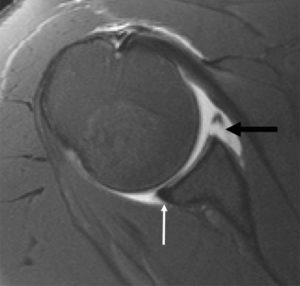 Hottya GA, Tirman PF, Bost FW, Montgomery WH, Wolf EM, Genant HK. in Radiology in 2008 examined 36 patients following acute traumatic shoulder dislocation and revealed full-thickness tears in 19% of patients and partial or full-thickness tears in 42%.17As would be expected, subscapularis tears were most common, but tears were also identified in the supraspinatus and the infraspinatus. 1992 Jul;74(6):890-6. 3.